Fundus Fluorescein Angiography (FFA), also known as Eye Angiography, is a medical imaging technique used to evaluate retinal diseases. It was first defined and applied by medical students H.R. Novotny and D.L. Alvis at Indiana University. The technique gained widespread acceptance in 1967 when Donald Gass shared his extensive research and experiences using FFA.
With advancements in computer software and digital imaging tools, the effectiveness and benefits of FFA have significantly improved.
For clarity and accessibility, the term Eye Angiography is often used interchangeably with Fundus Fluorescein Angiography in this article.
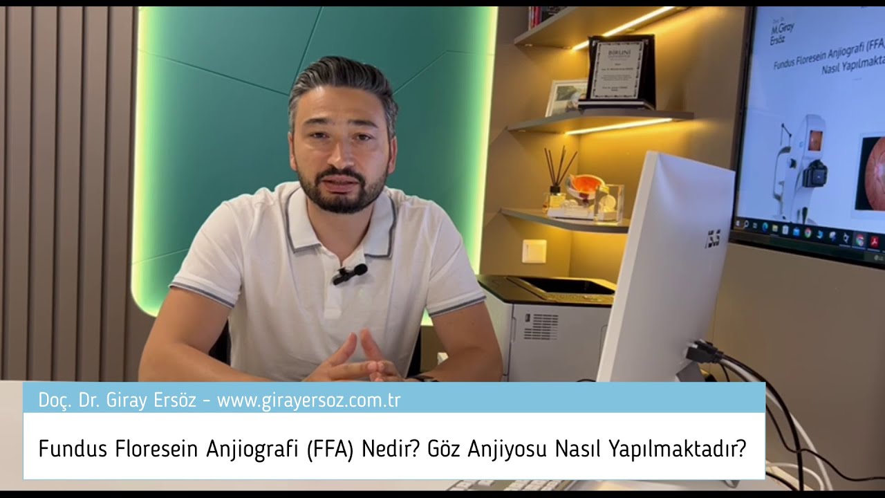
What is Fundus Fluorescein Angiography (FFA, Eye Angiography)?
The term “fundus” refers to the bottom part of organs that have a cavity in the middle, farthest from the entrance. In the context of the eye, the fundus includes the optic nerve, macula, retina, retinal blood vessels, and vitreous, which are located at the back of the eye. This area is often referred to as the “bottom of the eye” in Turkish.
Fundus Fluorescein Angiography (FFA), also known as Eye Angiography, is a technique primarily used to diagnose diseases affecting the back of the eye. FFA involves the use of a fluorescent dye called fluorescein to visualize the retinal vessels. This method is fundamental for diagnosing and monitoring retinal diseases.

How is Fundus Fluorescein Angiography (FFA, Eye Angiography) Performed?
Fundus Fluorescein Angiography (FFA), commonly known as eye angiography, is an imaging technique used to diagnose retinal diseases and assess their extent and severity. Unlike cardiac angiography, eye angiography is a non-invasive procedure that does not require hospitalization or the insertion of catheters.
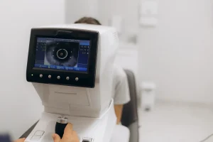
To perform FFA, a vascular access point is first established in the patient’s arm. A fluorescent dye called fluorescein is then injected through this access point. The patient remains seated while special filter-equipped cameras capture images of the retina. This process takes approximately 10 minutes, during which the patient may be asked to look in different directions to obtain comprehensive images of the retinal structures.
In Which Diseases Is Eye Angiography (Fundus Fluorescein Angiography, FFA) Used?
Fundus Fluorescein Angiography (FFA), or eye angiography, is widely used in diagnosing and monitoring various retinal diseases and uveitis. Key retinal conditions where FFA is particularly useful include:
- Age-related macular degeneration (AMD)
- Diabetic retinopathy
- Retinal vein and artery occlusions
- Central serous retinopathy (CSR, CSCR)
- Retinal vascular inflammation
- Macular edema
What are the Risks of Eye Angiography (Fundus Fluorescein Angiography, FFA)?
Fundus Fluorescein Angiography (FFA), or eye angiography, is considered a low-risk procedure among medical interventions. However, some side effects have been reported, with an overall incidence rate of approximately 0.083%. These side effects can be categorized by severity as mild (1.24%), moderate (0.2%), and severe (0.04%).
The most common side effects include:
- Nausea
- Vomiting
- Bitter taste
- Itching
- Abdominal pain
- Sneezing
In rare cases, individuals with a predisposition to allergies may experience serious allergic reactions. The fluorescein dye used in the procedure is eliminated from the body through the kidneys. Studies have shown that it does not cause significant deterioration in renal function, even in patients with kidney failure. Nonetheless, caution is advised when using FFA in patients with renal impairment and those on dialysis.
What is Indocyanine Green Angiography (ICGA)?
Indocyanine Green Angiography (ICGA) is another form of eye angiography that involves the intravenous administration of a dye called indocyanine green. While ICGA is less commonly used than Fundus Fluorescein Angiography (FFA) in retinal disease clinics, it is often employed in conjunction with FFA to confirm diagnoses in specific conditions.
ICGA is particularly useful in diagnosing certain types of age-related macular degeneration, such as polypoidal choroidal vasculopathy and retinal angiomatous proliferation. It is also valuable in assessing pachychoroid spectrum diseases, including central serous retinopathy and chorioretinitis.
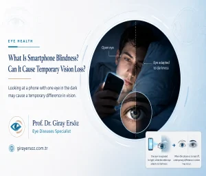
What Is Smartphone Blindness? Can It Cause Temporary Vision Loss?
Have you ever noticed short-term vision loss in one eye after looking at your phone [...]
Devamını OkuyunJul

How Can I Tell If My Eye Prescription Has Increased?
Your eye prescription may change over time. Sometimes this change is not noticed immediately; instead, [...]
Devamını OkuyunJul
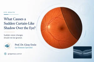
What Causes a Sudden Curtain-Like Shadow Over the Eye?
If you suddenly experience a darkening, shadowing, or closing sensation as if a curtain has [...]
Devamını OkuyunJul
How Is an Eye Examination Performed? When Should It Be Done in Babies and Children?
An eye examination is a medical evaluation that is vital for protecting eye health and [...]
Devamını OkuyunJul
What Is Hyperopia (Farsightedness)? Causes, Symptoms and Treatment
Hyperopia is one of the most common vision problems seen both in Türkiye and around [...]
Devamını OkuyunJul

What Is Myopia? What Are the Symptoms and Treatment Methods of Myopia?
Myopia, commonly known as nearsightedness, is a refractive error of the eye. People with myopia [...]
Devamını OkuyunJun

What Is Astigmatism? Symptoms and Treatment Methods for Astigmatism
Astigmatism is one of the most common refractive errors affecting millions of people today. It [...]
Devamını OkuyunJun

The Benefits of Wearing Sunglasses and How to Choose Them
Wearing sunglasses plays a vital role in protecting our eye health. However, choosing the wrong [...]
Devamını OkuyunSep