What Is Choroidal Hemangioma? Symptoms and Current Treatment
Choroidal hemangioma is one of the vascular tumoral lesions that can occur inside the eye. In this article, I answer common questions about the diagnosis, follow-up and treatment process of this condition.
What Is Choroidal Hemangioma?
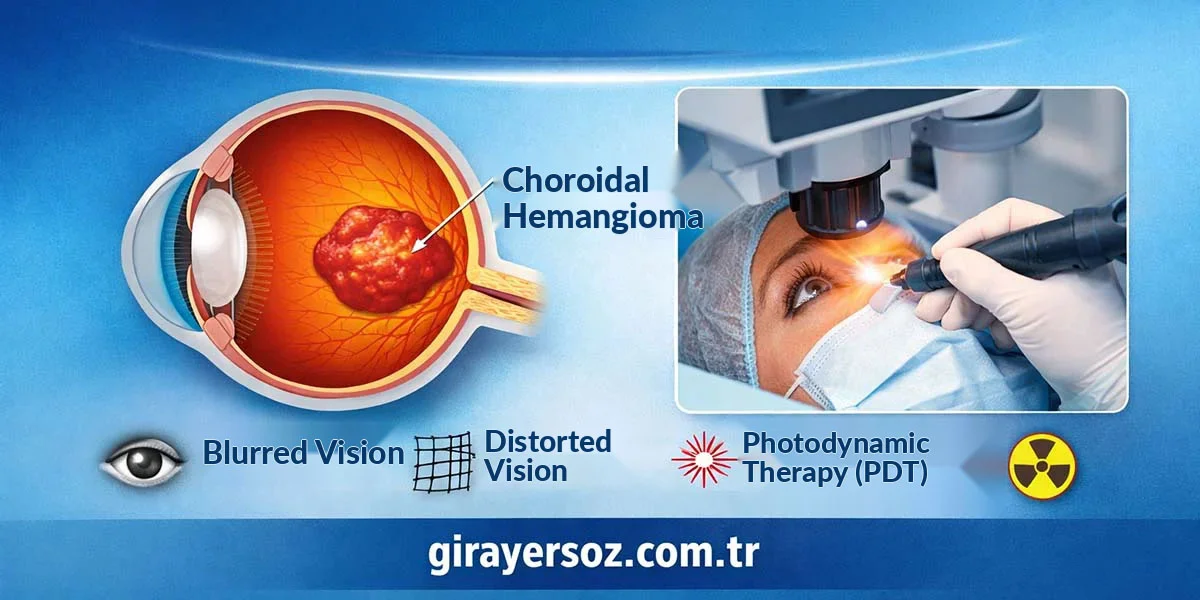
Choroidal hemangioma is a benign vascular tumour that originates from the choroid layer at the back of the eye. Choroidal hemangioma is not cancer.
The choroid is located between the retina, which contains the nerve cells responsible for vision, and the outermost layer of the eye. It has a dense network of blood vessels that nourish the retina. In choroidal hemangioma, these vessels become abnormally enlarged and form a sponge-like vascular mass. In some patients, this vascular structure may cause fluid to collect under the retina, leading to blurred or reduced vision.

Causes of Choroidal Hemangioma
Choroidal hemangioma usually develops due to congenital differences in vascular development. When the blood vessels in the choroid layer at the back of the eye are wider and denser than normal, this benign vascular mass may form. The most common type is circumscribed choroidal hemangioma. Circumscribed choroidal hemangioma usually affects only one eye, and in most patients, no clear genetic cause is identified. The rarer diffuse choroidal hemangioma is most often associated with Sturge-Weber syndrome. In this type, there are widespread vascular enlargements in the choroid, and patients may also have port-wine stain-like vascular marks on the face.
Sturge-Weber syndrome most commonly develops as a result of mutations in the GNAQ gene. This mutation is usually not inherited, meaning it is not passed from the mother or father; instead, it develops during the baby’s development in the womb. In the circumscribed type, the tumour margins can be clearly seen, while in the diffuse type, the tumour spreads over such a wide area that its borders cannot be clearly defined.
How Common Is Choroidal Hemangioma?
Choroidal hemangioma is a relatively rare vascular tumour of the eye. Current estimates suggest that its prevalence in the general population is approximately 0.0005% (about 1 in every 200,000 people). The vast majority of cases are circumscribed choroidal hemangiomas, which usually affect only one eye. The rarer diffuse choroidal hemangioma is most commonly associated with Sturge-Weber syndrome and accounts for only a small percentage of all choroidal hemangioma cases. Therefore, the exact rate of the diffuse type in the general population cannot be estimated.
Risk Factors for Choroidal Hemangioma
No risk factor has yet been clearly shown to contribute to the development of circumscribed choroidal hemangioma. For the rarer diffuse type, the most important risk factor is having Sturge-Weber syndrome.
Symptoms of Choroidal Hemangioma
Choroidal hemangioma may not cause any symptoms in the early stages. As tumours located close to the visual centre grow, the patient’s refractive value, in other words the glasses prescription, may shift from myopia toward hyperopia. When fluid leakage from the choroidal hemangioma begins under the retina, symptoms such as blurred vision, dark areas in vision, broken or distorted vision, and wavy vision may occur. After this stage, if choroidal hemangioma is not treated, permanent loss of visual acuity may begin to develop.
Diagnosis of Choroidal Hemangioma
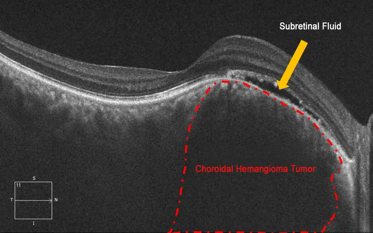
To diagnose choroidal hemangioma, a dilated fundus examination must first be performed. During the examination, this tumour appears under the retina as an elevated red-orange nodular structure. The diagnosis is then confirmed with OCT (optical coherence tomography), fluorescein angiography and indocyanine green angiography tests. In atypical-looking cases that need to be differentiated from other tumours or cancers, ultrasonography and MRI may also be performed.
Treatment Methods for Choroidal Hemangioma
The treatment method for choroidal hemangioma is chosen by evaluating the stage of the disease and its effect on eye health.
Observation
Small choroidal hemangiomas that do not cause symptoms and have not caused fluid leakage under the retina can be monitored at regular intervals with vision examinations and OCT testing.
Photodynamic Therapy (PDT)
The most current and gold-standard treatment for choroidal hemangioma is photodynamic therapy. In choroidal hemangiomas that cause leakage, photodynamic therapy is the most commonly used method because it is highly effective and carries a lower risk of damaging the surrounding tissue.
For detailed information about photodynamic therapy, you can read our photodynamic therapy article.
Laser Photocoagulation / Transpupillary Thermotherapy
Transpupillary thermotherapy laser treatment (TTT) is an effective treatment method for choroidal hemangioma. However, because it carries a higher risk of thermal damage to the retina over the tumour and the surrounding tissues, it can only be used for tumours located away from the visual centre.
Radiotherapy
Plaque radiotherapy and external beam radiotherapy may be used to reduce tumour size and leakage in very large choroidal hemangiomas or in diffuse choroidal hemangiomas associated with Sturge-Weber syndrome. However, it should be known that radiotherapy carries a risk of radiation retinopathy (damage to the retinal blood vessels) or optic neuropathy (damage to the optic nerve).
Intraocular Injections
Intravitreal anti-VEGF injections administered into the eye do not have a direct effect on tumour size. However, they may be used to reduce leakage in patients for whom other treatments cannot be applied, or as an additional treatment alongside other methods.
Source
- Circumscribed Choroidal Hemangioma
- Circumscribed choroidal hemangioma: An overview of clinical manifestation, diagnosis and management
- Circumscribed Choroidal Hemangiomas
- Photodynamic therapy of circumscribed choroidal haemangioma
- Circumscribed choroidal hemangioma: Clinical manifestations and factors predictive of visual outcome in 200 consecutive cases
- Incidence of Sturge-Weber Syndrome and Associated Ocular Involvement
- Current concepts on diffuse choroidal hemangioma in Sturge-Weber syndrome
- Choroidal hemangioma in Sturge-Weber syndrome
Frequently Asked Questions (FAQ)
Choroidal hemangioma is not cancer.
If choroidal hemangioma is not treated, it can cause the retinal cells responsible for vision in the affected area to die. Therefore, if tumours affecting the central visual area, known as the macula, are not treated, they may lead to permanent loss of central vision.
Patients treated with photodynamic therapy (PDT) at an early stage may regain 100% vision. However, the longer treatment is delayed, the greater the risk of permanent vision loss.
Choroidal hemangioma is a tumour that can recur. In various clinical series, the recurrence rate has been reported as 10–20%.
Choroidal hemangioma very rarely needs to be surgically removed. The safest treatment is photodynamic therapy.
Choroidal hemangioma does not prevent exercise.

How Can I Tell If My Eye Prescription Has Increased?
Your eye prescription may change over time. Sometimes this change is not noticed immediately; instead, [...]
Devamını OkuyunJul
What Causes a Sudden Curtain-Like Shadow Over the Eye?
If you suddenly experience a darkening, shadowing, or closing sensation as if a curtain has [...]
Devamını OkuyunJul
How Is an Eye Examination Performed? When Should It Be Done in Babies and Children?
An eye examination is a medical evaluation that is vital for protecting eye health and [...]
Devamını OkuyunJul

What Is Hyperopia (Farsightedness)? Causes, Symptoms and Treatment
Hyperopia is one of the most common vision problems seen both in Türkiye and around [...]
Devamını OkuyunJul

What Is Myopia? What Are the Symptoms and Treatment Methods of Myopia?
Myopia, commonly known as nearsightedness, is a refractive error of the eye. People with myopia [...]
Devamını OkuyunJun
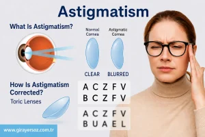
What Is Astigmatism? Symptoms and Treatment Methods for Astigmatism
Astigmatism is one of the most common refractive errors affecting millions of people today. It [...]
Devamını OkuyunJun

The Benefits of Wearing Sunglasses and How to Choose Them
Wearing sunglasses plays a vital role in protecting our eye health. However, choosing the wrong [...]
Devamını OkuyunSep

The Effects of Eyelash Trimming on Eye Health
Trimming eyelashes has recently gained popularity on social media, especially among men. While it may [...]
Devamını OkuyunSep