Understanding the anatomy of the eye is crucial for diagnosing and treating various eye conditions. Additionally, eye exams can often provide early detection of systemic diseases, such as diabetes and hypertension.
İçindekiler
What is the Eye?
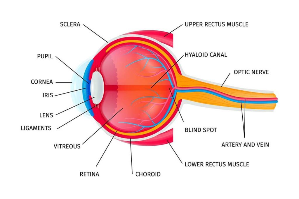
The eye is a vital sensory organ in the human body, responsible for capturing light and transmitting visual information to the brain. Composed of structures such as the iris, pupil, lens, and retina, the eye works in harmony to process images. Light first enters through the cornea and pupil, reaching the lens. The lens refracts the light and flips the image. This image is then projected onto the retina, where light is converted into electrical signals. These signals travel to the brain via the optic nerve, where they are interpreted and corrected to form a clear image. Additionally, the eye plays a key role in recognizing emotions and facilitating non-verbal communication.

The Structure and Functions of the Eye
The eye is a complex organ, and each of its structures plays a unique role in vision and overall eye health. The following outlines the main structures of the eye, their functions, and common diseases associated with them:
Cornea
As the outermost layer of the eye, the cornea serves as the first point of refraction for incoming light. Its transparent, durable structure protects the eye from infections. The cornea is composed of dense type 1 and type 5 collagen fibrils, which contribute to its strength and transparency through their specialized arrangement.
Corneal infections, or keratitis, are rare but can occur due to conditions such as chronic eyelash root inflammation, trauma, improper contact lens use, or excessive use of cortisone eye drops. Keratoconus is another significant condition where part of the cornea becomes steeper and protrudes in a cone shape, causing thinning in that area.
Sclera (Hard Layer)
The sclera, or the white part of the eye, provides structural support and protection to the eyeball. It also serves as the attachment point for the eye muscles. While the sclera is similar to the cornea in its composition, it is not transparent due to the different arrangement of its collagen fibers. It is made mostly of type 1 collagen, with some type 3 collagen.
Scleral diseases are rare, but scleritis, which is inflammation of the sclera, can occur, often associated with autoimmune or rheumatic diseases.
Conjunctiva
The conjunctiva is the thin, transparent membrane that covers the inner surface of the eyelids and the white part of the sclera. When inflamed, typically due to infection or allergies, this condition is known as conjunctivitis. Conjunctivitis is a common cause of red eyes.
Iris
The iris is the colored part of the eye, with the pupil (the central opening) controlling the amount of light entering the eye. The iris, along with the ciliary body and choroid, forms the uveal tissue. Inflammation of the uveal tissues is referred to as uveitis and when the iris alone is affected, it is known as anterior uveitis.
Vascular Reticular Layer (Choroid)
The choroid lies beneath the sclera and above the retina, and it contains blood vessels that provide oxygen and nutrients to the eye. The outer two-thirds of the retina rely on the choroid for nourishment, meaning that diseases of the choroid often affect the retina as well. Some of the most common choroidal diseases include central serous chorioretinopathy (CSC), choroidal neovascularization (CNV) in wet age-related macular degeneration, and chorioretinitis, which is inflammation of both the choroid and retina.
Retina
The retina is a crucial tissue located at the inner part of the eye, responsible for vision. It is attached to the vitreous gel inside the eye and is supported by the choroid layer on the outside. The retina contains photoreceptor cells, which are essential for visual perception. These cells come in two types: rods and cones. Rods are responsible for vision in low light conditions, while cones enable color vision and sharp focus. The retina constitutes approximately 70% of the eye’s cellular structure, capturing light and forming images. Additionally, it converts light into nerve signals, which are transmitted to the brain through the optic nerve. A key area of the retina is the macula, which will be discussed separately.
Diseases affecting the retina include macular degeneration, retinal tears, and rhegmatogenous retinal detachment. Macular degeneration specifically affects the macula, leading to a decline in vision.
Macular Degeneration
The macula is a small, vital area at the center of the retina responsible for sharp, detailed vision. Known medically as the macula lutea, it contains cone cells that provide the clearest vision and enable color perception. The macula is roughly 5.5 mm in diameter, with the fovea in its center being the region that allows the sharpest vision. The fovea itself is about 0.35 mm in diameter.
The primary diseases associated with macular degeneration include age-related macular degeneration (AMD), diabetic macular edema, macular degeneration due to retinal vein occlusion, central serous retinopathy (CSR), macular holes, and epiretinal membrane (ERM) formation.
Vitreous
The vitreous is a transparent, gel-like substance located inside the eye. It helps maintain the eye’s shape and assists in refracting light. The vitreous is attached to the retina and typically begins to separate from it as a person ages. While this separation generally occurs gradually, it can sometimes lead to retinal tears or macular holes.
Other conditions affecting the vitreous include vitreous opacities, which can lead to floaters, and vitreous hemorrhages, which involve bleeding within the vitreous.
Optic Nerve
The optic nerve is the largest nerve in the eye. It is formed by the extensions of nerve cells in the retina and carries the electrical signals generated by these cells to the brain, effectively transmitting the visual image from the eye. Damage to the optic nerve can lead to vision loss.
Key conditions affecting the optic nerve include optic neuritis, anterior ischemic optic neuropathy, and papilledema.
Lens (Eye Lens)
The lens is a transparent structure located behind the eye that refracts light to help focus vision. Much like a camera lens, it adjusts the focus for clear vision. Over time, the lens can become less flexible, particularly after the age of 40, causing difficulty with near vision. A common condition related to the lens is cataracts, which cause the lens to lose transparency, leading to vision problems.
The Benefits of Wearing Sunglasses and How to Choose Them
Wearing sunglasses plays a vital role in protecting our eye health. However, choosing the wrong [...]
Devamını OkuyunSep
The Effects of Eyelash Trimming on Eye Health
Trimming eyelashes has recently gained popularity on social media, especially among men. While it may [...]
Devamını OkuyunSep
The Benefits of the Mediterranean Diet for Macular Degeneration
Diet plays a significant role in slowing the progression of macular degeneration, and the Mediterranean [...]
Devamını OkuyunApr
What is Dry Eye? Tips to Protect Yourself from Dry Eyes
Insufficient tear production can lead to discomfort and reduce your eyes’ ability to defend against [...]
Devamını OkuyunJan
Understanding the Structure of the Eye and Its Functions?
Understanding the anatomy of the eye is crucial for diagnosing and treating various eye conditions. [...]
Devamını OkuyunJan
Causes and Treatment of Eye Floaters
Occasionally, individuals may notice floating objects in their field of vision. These can take various [...]
Devamını OkuyunMay
What is Vitreous? How is Vitreous Dysfunction Treated?
Vitreous, derived from the Latin term meaning “glassy,” is a clear gel found between the [...]
Devamını OkuyunMay
Vision Wellness Essentials: Simple and Effective Tips for Eye Health
Our eyes, vital and health-sensitive organs, demand special care to combat conditions such as eye [...]
Devamını OkuyunJan