Various tests are conducted to detect, diagnose, and monitor the progression of eye diseases. One such test is visual field testing, which is commonly used to evaluate conditions like glaucoma, macular degeneration, and retinitis pigmentosa.
In this article, I’ll begin by explaining what the visual field is and then provide a detailed overview of visual field testing.
What is the Visual Field?
The visual field refers to the entire area a person can see when looking straight ahead. The extent of the visual field varies across different regions. For instance, the field of vision near the nose is about 60º, while the area beneath the eyes covers roughly 70º. The upper visual field extends around 50º, and the field on the side of the ear reaches up to 90º. This wider range on the ear side is due to the absence of any obstruction from facial structures.
The visual field can be categorized into two types: central vision (the area directly in front of the observer) and peripheral vision (the outer or side vision).
What is a Visual Field Test?
A visual field test is a psychophysical assessment used to detect any loss in a person’s central or peripheral vision. If a loss is present, the test also helps determine the extent of this vision impairment. While this test is commonly used for a variety of eye conditions, it is especially important for diagnosing glaucoma.
Why is a Visual Field Test Performed?
A visual field test is conducted for several reasons. Primarily, it helps identify eye diseases, particularly glaucoma and certain retinal disorders. Additionally, the test is crucial during the treatment process, as it allows doctors to monitor disease progression and assess whether the condition is worsening.
The visual field test is also crucial in differentiating between optic disc drusen and papilledema, two conditions that can sometimes be confused. By using this test, misdiagnoses can be avoided. It is also valuable for diagnosing and monitoring conditions like night blindness (Chicken Black) and tracking the progress and recovery of the disease. Additionally, visual field testing may be necessary for detecting neurological issues, including optic neuritis, brain tumors, and strokes.
Traditional Visual Field Tests
In addition to computerized methods, visual field assessments can also be carried out using traditional techniques. Some of the most widely used traditional visual field tests include the Amsler Card and Confrontation test, which are outlined below.
Amsler Card Visual Field Test Application
One of the traditional methods involves using Amsler Cards. These cards are 10 cm in length and feature a grid of 5 mm squares. There is a central point on the card that the patient should focus on. While concentrating on this central point, the patient is asked about any irregularities they notice, such as blurred squares or areas where squares are missing entirely. The patient then reports how they perceive other parts of the card. The Amsler card is primarily used to assess the central visual field and is particularly helpful in diagnosing conditions affecting the macula, such as age-related macular degeneration (AMD).
Confrontation is another traditional test used to evaluate the visual field.
Confrontation Visual Field Test Application
In the confrontation test, the individual with normal vision sits face-to-face with the person suspected of having a visual field issue. During the test, the patient closes their right eye while the examiner closes their left eye. The examiner then slowly moves an object towards the patient’s visual field from different angles to determine at what point the patient perceives it. This process is repeated for the other eye as well.
Another traditional method, known as the “curtain test“, involves assessing whether the patient notices light reflections on a curtain. The light is varied in intensity, alternating between brighter and dimmer levels, to evaluate the patient’s ability to detect changes in the visual field.
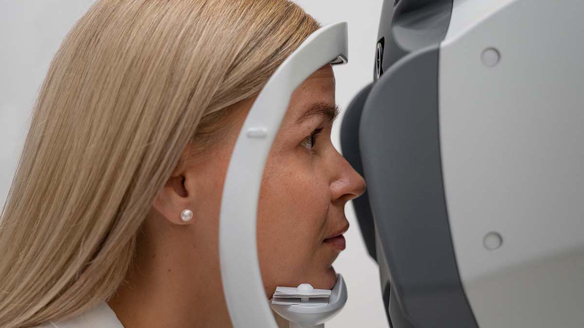
Modern Visual Field Testing Procedure
Currently, visual field testing is conducted using specialized devices designed for this purpose. Separate tests are performed for each eye. The patient positions their head on the apparatus in front of the device, resting their chin securely. While keeping one eye closed, the patient focuses on a light located on the opposite side. During the test, flashes of light, both weak and intense, appear at various locations in the patient’s peripheral vision. The patient is instructed to press a button whenever they detect a flash, indicating their ability to perceive the light. This process helps assess the patient’s sensitivity to light and determines how much their retinal sensitivity deviates from normal levels.

The Benefits of Wearing Sunglasses and How to Choose Them
Wearing sunglasses plays a vital role in protecting our eye health. However, choosing the wrong [...]
Devamını OkuyunSep
The Effects of Eyelash Trimming on Eye Health
Trimming eyelashes has recently gained popularity on social media, especially among men. While it may [...]
Devamını OkuyunSep

The Benefits of the Mediterranean Diet for Macular Degeneration
Diet plays a significant role in slowing the progression of macular degeneration, and the Mediterranean [...]
Devamını OkuyunApr

What is Dry Eye? Tips to Protect Yourself from Dry Eyes
Insufficient tear production can lead to discomfort and reduce your eyes’ ability to defend against [...]
Devamını OkuyunJan

Understanding the Structure of the Eye and Its Functions?
Understanding the anatomy of the eye is crucial for diagnosing and treating various eye conditions. [...]
Devamını OkuyunJan

Causes and Treatment of Eye Floaters
Occasionally, individuals may notice floating objects in their field of vision. These can take various [...]
Devamını OkuyunMay

What is Vitreous? How is Vitreous Dysfunction Treated?
Vitreous, derived from the Latin term meaning “glassy,” is a clear gel found between the [...]
Devamını OkuyunMay

Vision Wellness Essentials: Simple and Effective Tips for Eye Health
Our eyes, vital and health-sensitive organs, demand special care to combat conditions such as eye [...]
Devamını OkuyunJan