Various medical devices are used during eye examinations to diagnose eye diseases. Optical Coherence Tomography (OCT), commonly referred to as eye tomography, is one of the most frequently utilized devices. OCT provides detailed information about the structure of the eyes and the presence of eye diseases.
The abbreviation for Optical Coherence Tomography is OCT.
What is Optical Coherence Tomography (OCT – Eye Tomography)?
Optical Coherence Tomography (OCT), also known as Eye Tomography, is an advanced imaging technique used to capture detailed cross-sectional images of the retina, the light-sensitive layer at the back of the eye. By using light waves, OCT provides high-resolution images of all retinal layers, allowing for precise mapping and measurement of retinal thickness.
These detailed images and measurements are invaluable in diagnosing retinal diseases and guiding ophthalmologists in the treatment process.
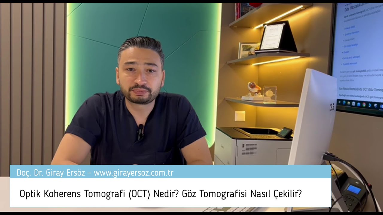
How is Optical Coherence Tomography (OCT – Eye Tomography) Performed?
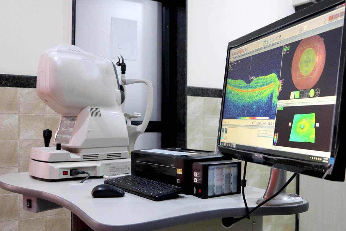
Optical Coherence Tomography (OCT), also known as Eye Tomography, is typically conducted by trained technicians or nurses. In many cases, pupil dilation is not required for the procedure. The patient positions their chin on the device, and the technician performs the measurements, with each scan taking approximately 10-20 seconds. Detailed images are captured within 1-2 minutes, providing valuable information for diagnosing retinal diseases, glaucoma, and optic nerve conditions. Subsequently, the doctor develops a treatment plan based on these findings.
Which Eye Diseases are Diagnosed Using Optical Coherence Tomography (OCT – Eye Tomography)?
Optical Coherence Tomography (OCT), also known as Eye Tomography, plays a crucial role in diagnosing various eye conditions, including:
- Macular hole
- Epiretinal membrane
- Macular edema
- Macular degeneration
- Glaucoma
- Central serous retinopathy
- Diabetic retinopathy
Additionally, OCT is utilized to assess disorders affecting the optic nerve, which transmits visual signals from the retina to the brain.
Why is OCT (Eye Tomography) Used in Macular Degeneration?
OCT (Eye Tomography) is essential for diagnosing age-related macular degeneration. It provides high-resolution cross-sectional images of the retina, revealing its morphological structure without the need for invasive procedures or medication. This non-invasive method ensures patient safety as it does not require vascular access.
Eye tomography allows for clear identification of bleeding or edema beneath the macula, which is crucial for diagnosing macular degeneration.
Why is OCT (Eye Tomography) Used in Glaucoma?
Glaucoma, a progressive eye condition often associated with elevated intraocular pressure, can lead to vision loss if left untreated. Optical Coherence Tomography (OCT), or eye tomography, plays a critical role in diagnosing this disorder.
OCT is particularly valuable because it can detect as little as 1-2% of optic nerve damage years before visual field loss occurs. Early detection is crucial in managing glaucoma effectively, similar to other diseases. It helps in accurate diagnosis, preventing misdiagnosis and inappropriate medication use. Regular OCT scans also provide vital insights to the doctor regarding the disease’s progression.
Why is OCT (Eye Tomography) Used in Macular Hole?
Optical Coherence Tomography (OCT), or eye tomography, is instrumental in diagnosing macular holes. To diagnose a macular hole, the pupil is typically dilated using eye drops, facilitating clearer imaging of the retina with OCT. This non-invasive procedure uses light waves to create detailed images of the retina.
After OCT imaging, which is safe and devoid of side effects, the presence and severity of a macular hole are determined. This information guides the ophthalmologist in prescribing the appropriate treatment for the patient.
What are the Conditions that May Occur After Optical Coherence Tomography (OCT – Eye Tomography)?
Optical Coherence Tomography (OCT), or eye tomography, typically does not cause side effects. However, in cases where pupil dilation is required using drops (drop tomography), some individuals may experience temporary eye redness and a sensation of burning.
If short-acting drops are used, blurry vision may occur within 6 hours after the procedure, while with long-acting drops, blurry vision may persist for up to 24 hours. Despite these possible effects, eye tomography is generally comparable to undergoing routine photography and is well-tolerated under normal conditions.
Frequently Asked Questions About Optical Coherence Tomography (OCT – Eye Tomography)
Are There Any Harms of Optical Coherence Tomography (OCT – Eye Tomography)?
Eye tomography involves capturing images of the eye using reflected light, without the use of radiation. This non-invasive procedure poses no harm to patients and is safe for diagnostic purposes.
Who Receives Optical Coherence Tomography (OCT – Eye Tomography)?
Optical Coherence Tomography (OCT – Eye Tomography) can be performed on individuals of all ages with various eye conditions. It is a versatile diagnostic tool utilized from pediatric to elderly patients. However, tomography is typically postponed in patients with eye infections to prevent inaccurate measurements and the potential spread of infection to others.
Are Optical Coherence Tomography (OCT – Eye Tomography) Results Available Immediately?
Yes, the results of optical coherence tomography are available immediately after the procedure. Following the imaging, a doctor will interpret the results and proceed with diagnosis and treatment planning.

The Benefits of Wearing Sunglasses and How to Choose Them
Wearing sunglasses plays a vital role in protecting our eye health. However, choosing the wrong [...]
Devamını OkuyunSep

The Effects of Eyelash Trimming on Eye Health
Trimming eyelashes has recently gained popularity on social media, especially among men. While it may [...]
Devamını OkuyunSep

The Benefits of the Mediterranean Diet for Macular Degeneration
Diet plays a significant role in slowing the progression of macular degeneration, and the Mediterranean [...]
Devamını OkuyunApr

What is Dry Eye? Tips to Protect Yourself from Dry Eyes
Insufficient tear production can lead to discomfort and reduce your eyes’ ability to defend against [...]
Devamını OkuyunJan

Understanding the Structure of the Eye and Its Functions?
Understanding the anatomy of the eye is crucial for diagnosing and treating various eye conditions. [...]
Devamını OkuyunJan

Causes and Treatment of Eye Floaters
Occasionally, individuals may notice floating objects in their field of vision. These can take various [...]
Devamını OkuyunMay

What is Vitreous? How is Vitreous Dysfunction Treated?
Vitreous, derived from the Latin term meaning “glassy,” is a clear gel found between the [...]
Devamını OkuyunMay

Vision Wellness Essentials: Simple and Effective Tips for Eye Health
Our eyes, vital and health-sensitive organs, demand special care to combat conditions such as eye [...]
Devamını OkuyunJan